Figure 5.
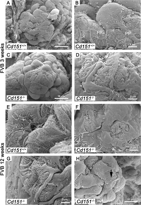
SEM Analysis of Podocyte Structure in FVB Cd151-Null Mice. Three-and 12-week old FVB Cd151+/+ and Cd151−/− kidneys were analyzed by SEM to determine the extent of glomerular and more specifically podocyte damage. In wild-type animals (A, B, and E) the podocyte structure is well defined with the cell body dividing into primary processes (PP) that divide into foot processes (FP). At 3 weeks of age, the podocytes look abnormal in Cd151−/− kidneys (C, D) compared to wild-type controls (A, B). Indeed, the podocyte cell bodies look flattened at 3 weeks of age (C). Primary processes and foot processes are still present in most areas as seen by TEM but appear disorganized (C, D, and G). Podocyte alterations progress with age and foot process effacement has become more pronounced by 12 weeks (F). However, even at 12 weeks of age, there are still a number of places where disorganized foot processes are still visible (G). Also note the unhealthy podocyte sending a process onto the cell body of its neighbor (arrow in H). The representative results from 3 mice in each group at 3 weeks of age and two mice in each group at 12 weeks of age are shown. Three to 6 glomeruli were analyzed for each mouse. Original magnification: ×6000 (A, C); ×20,000 (B, D–G); ×12,000 (H).

