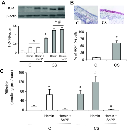
Figure 1.
HO-1 expression and HO activity in rats exposed or not to CS. A: Typical Western blot showing detection of HO-1 protein expression. The bottom part shows densitometric analysis of HO-1 expression expressed as a ratio of β-actin expression. C, Exposure to room air; CS, exposure to cigarette smoke; H, hemin; SnPP-IX, tin protoporphyrin IX. Values are mean ± SEM, n = 8 to 10 in each group. *P < 0.05 versus vehicle-treated C; #P < 0.05 versus vehicle-treated CS. B: Immunohistochemical analysis of HO-1 expression in the trachea of one rat exposed to room air (C) and in another exposed to cigarette smoke (CS). Staining was observed in the tracheal epithelium of the CS rat. No staining was observed with the isotype antibody (data not shown). The bottom part shows the percentage of HO-1 (+) cells over the whole number of epithelial cells. Values are mean ± SEM, n = 8 to 10 in each group. *P < 0.05 versus C. C: HO activity. C, Exposure to room air; CS, exposure to cigarette smoke; H, hemin; SnPP-IX, tin protoporphyrin IX. Abbreviations are the same as those in A. Values are mean ± SEM, n = 8 to 10 in each group. *P < 0.05 versus vehicle-treated C; #P < 0.05 versus vehicle-treated CS. Original magnifications, ×40.

