Figure 3.
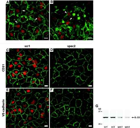
Inhibition of the junctional protein VE-cadherin does not alter the expression level of IL-33 and knockdown of IL-33 does not alter the cellular distribution or expression level of junctional proteins CD31 or VE-cadherin. Immunocytochemical double staining for IL-33 (IL-33Nter, red) and VE-cadherin (mouse IgG1, green) in HUVECs preincubated with vehicle (PBS, A) or anti-VE-cadherin (IgG2a, B) from point of seeding until fixation. Pictures were obtained using identical exposure times and image-enhancement parameters. Arrowheads point to areas of intercellular contact. Note that cells in B with redistribution or lowered expression level of VE-cadherin maintained expression of IL-33. C and D: Immunocytochemical double staining for IL-33 (IL-33Nter, red) and CD31 (green) in HUVECs transfected with mock (C) or specific siRNA (D). E and F: Immunocytochemical double staining for IL-33 (red) and VE-cadherin (green) in HUVECs transfected with mock (E) or specific siRNA (F). Pictures were obtained using identical exposure times and image enhancement parameters. G: Western blot of IL-33 in cell lysates of HUVEC monolayers transfected with either scrambled siRNA controls (scr1 and scr2) or concentration-matched IL-33 targeting siRNA (spec1 and spec2). Shown are lanes from the same experiment, which was independently repeated twice with similar results. Scale bars = 10 μm.

