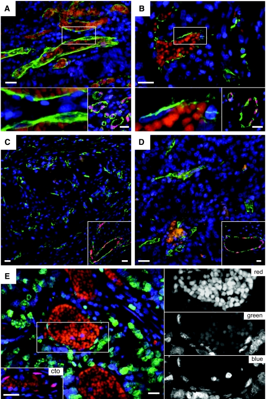
Figure 4.
Vascular IL-33 expression is lost in tumor vessels. Endothelial cells in vessels of human colon adenocarcinoma (A), ductal mammary carcinoma (B), renal clear cell carcinoma (C), and renal papillary carcinoma (D) were stained for IL-33 (Nessy-1, red) and with Ulex europaeus lectin (green) and Hoechst dye (blue) to visualize endothelial cells and cell nuclei, respectively. (For consistency throughout the figures, the red and green channels in Figure 4 were reversed.) The bottom left panels in A and B show high-power magnifications of the respective boxed areas. Note presence of autofluorescent intravascular red blood cells in A and B as well as lack of IL-33 signal in endothelial cell nuclei. The bottom right panels in A to D and bottom left panel in E show IL-33-positive vessels in tissue surrounding the tumor in the same section. E: Human colon adenocarcinoma (different sample from A) was stained for IL-33 (Nessy-1, red), Ki-67 (green), and Hoechst dye (blue). Autofluorescent erythrocytes indicate blood vessels. Right: High-power magnifications of the boxed area in E separating the different colors. Note the lack of IL-33 signal in Ki-67-positive endothelial nuclei. Scale bars = 50 μm.

