Figure 6.
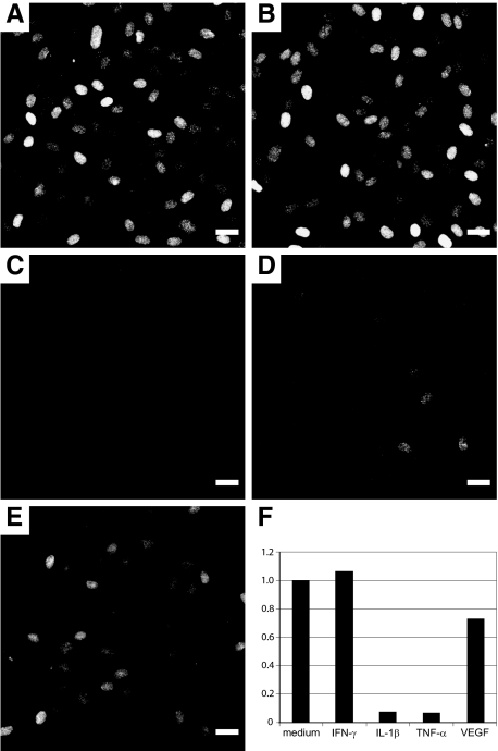
IL-33 expression is down-regulated by proinflammatory cytokines and VEGF. A--E: Response of superconfluent HUVEC monolayers exposed for 17 hours to medium alone (A), INF-γ (50 ng/ml) (B), IL-1β (0.5 ng/ml) (C), TNF-α (0.5 ng/ml) (D), or VEGF (10 ng/ml) (E). Immunostaining for IL-33 (IL-33Nter) after fixation of monolayers. Pictures were obtained using identical exposure times and image enhancement parameters. F: Quantitative PCR analysis of IL-33 expression in superconfluent HUVEC monolayers treated with cytokines for 7 hours at the same concentrations as given above. IL-33 transcript levels were obtained using primer pair IL-33 no. 2 and normalized to HPRT, data are given as ratio of transcript values in sample over those in medium-treated controls. Similar results were obtained when using a different primer pair for IL-33 detection or when normalizing against housekeeping gene GAPDH (data not shown). Scale bars = 25 μm.

