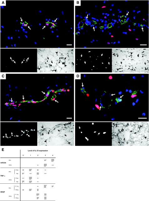
Figure 8.
TNF-α and VEGF down-regulate IL-33 in vivo. Immunostaining for IL-33 (Nessy-1, red) and CD31 (green) of rat skin after subcutaneous injection of PBS (A, C) and cytokines (B, D). Cell nuclei are stained with Hoechst dye. The bottom left panels show the red channel and the bottom right panels the light microscopy images of the respective area. Twenty-four hours after the injection of 0.4 μg of rrTNF-α (B) or 8 hours after the injection of 100 ng of rrVEGF (D) the endothelial signal for IL-33 has decreased compared to the mock injection (A and C, respectively). Pictures were obtained using identical exposure times and image enhancement parameters for A and B as well as for C and D. Arrows indicate endothelial nuclei. Arrowheads indicate ink particles. E: Semiquantitative analysis of vascular IL-33 expression in rat skin after injection of vehicle (PBS), rrTNF-α, or rrVEGF. Endothelial cell nuclei were categorized as negative (0) or increasingly positive (levels 1 to 4) by a blinded observer, counting in each condition 20 nuclei. Scale bars = 50 μm.

