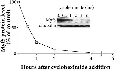
Figure 2.
Myf5 has a short half-life. C2 cells were cultured for 48 h in proliferation medium before being treated with 15 μg/ml CHX added to the medium. Myf5 and α-tubulin protein levels were followed by immunoblot analysis at the indicated time after CHX addition. Immunoblots were quantified by densitometric scanning, and Myf5 protein levels were expressed as the ratio of Myf5/α-tubulin signals.

