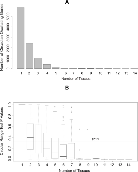
Figure 1. Tissue distribution of circadian oscillating genes.
(A) Distribution of the number of circadian oscillating genes identified in different numbers of mouse tissues. (B) Distribution of p-values in circular range tests for circadian phases of circadian oscillating genes identified in different numbers of mouse tissues.

