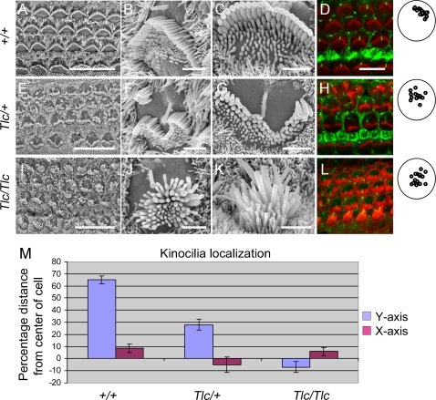
Figure 3. The kinocilia is mislocalized in Tailchaser hair cells.
(A–I) Scanning electron microscopy images showing auditory sensory epithelia of wild type (A–C), Tlc/+ (D–F) and Tlc/Tlc (G–I) mice at P0. Low magnification images (A, D and G) show that the characteristic arrangement of three rows of outer hair cells and a single row of inner hair cells is maintained in Tailchaser mutants. In wild type animals, outer (B) and inner (C) hair cell stereocilia form already highly organized bundles with kinocilium present at the center of the apical plane (arrow). Tailchaser mutants, however, show highly disorganized and misshaped stereocilia bundles on outer (E and H) and inner hair cells (F and I). Also, the position of the kinocilium was highly variable in Tailchaser mutants. Note remnants of a staircase-like stereocilia arrangement in Tlc/+ hair cell. (D, H, L) Confocal microscopy images showing kinocilium position in wild type (D), Tlc/+ (H) and Tlc/Tlc (L) at P0. Actin in stereocilia bundles was labeled with phalloidin (red). The kinocilia are labeled with an antibody that detects acetylated tubulin (green). A summary of positions of the kinocilia in outer hair cells demonstrates that kinocilia are more centrally located in mutants. (M) Kinocilium localization is represented as percentage of the distance from the center of the cell to its perimeter both in the Y (modiolar-striolar) and in the X (apical-basal) axes. Scale bars: A, D and G–15 µm; B, C, E, F, H, I–2 µm; D, H, L–5 µm.

