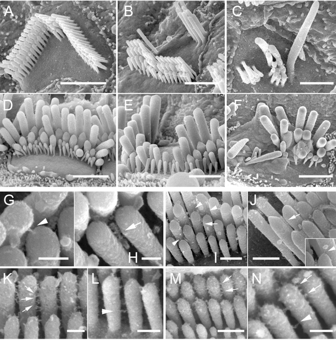
Figure 4. The Tailchaser mutation does not affect formation of interstereocilial links.
High resolution SEM images showing stereocilia bundles on outer (A–C, G–N) and inner (D–F) hair cells of wild type (A, D, G, H, K and L), Tlc/+ (B, E, I, M) and Tlc/Tlc (C, F, J and N) mice at P21 (G–J) and P7 (K–N). Note misshaped but staircase-like stereocilia bundles on hair cells of Tlc/+ animals (B and E) and disorganized bundles formed from fewer stereocilia of Tlc/Tlc mice (C and F). The horizontal connectors, tip links (G–J, tip links indicated by arrowheads, horizontal connectors indicated by arrows, inset on J shows high magnification image of tip link found between Tlc/Tlc stereocilia), lateral and ankle links (K–N, lateral links indicated by arrows, ankle links indicated by arrowheads) are present between Tlc/+ and Tlc/Tlc stereocilia and are indistinguishable from those of wild type mice. Scale bars: A–F-2 µm; G, H, K, L, N–200 nm; I, J and M–500 nm.

