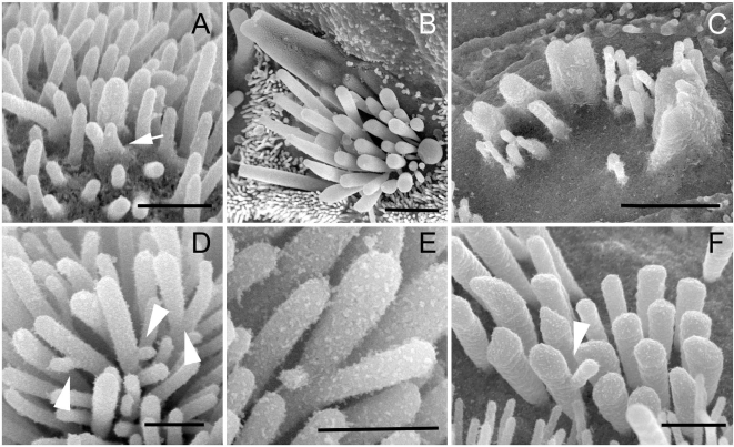
Figure 5. Stereocilium branching is observed in myosin VI mouse mutant hair bundles.
(A–C) High magnification SEM images showing stereocilia fusion in developing outer hair cells of Tlc/Tlc mice at P1 (A, apical turn) and inner (B, apical turn) and outer (C, basal turn) hair cells of Tlc/Tlc mice at P21. Note that stereocilia fusion starts at base of the stereocilia. (D–F) SEM images showing unusual branches found on Tailchaser stereocilia at P1 (D and E) and Snell's waltzer stereocilia at P6. Scale bars: A, D–F–500 nm; B and C–2 µm.

