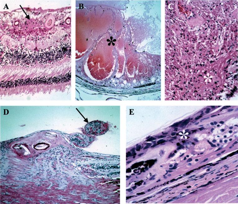
Figure 1.

Retinal hamartomas. (A) A small vascular hamartoma (arrow) located in the inner retina. (B) A large vascular hamartoma (asterisk) infiltrated most of the retinal layers. (C) A glial hamartoma in the retina (asterisk). (D) Two small hamartomas (arrow) located at the optic nerve head. (E) CPERH (asterisk) (haematoxylin and eosin; original magnification: A, ×200; B, ×50; C, ×200; D, ×100; E, ×400)
