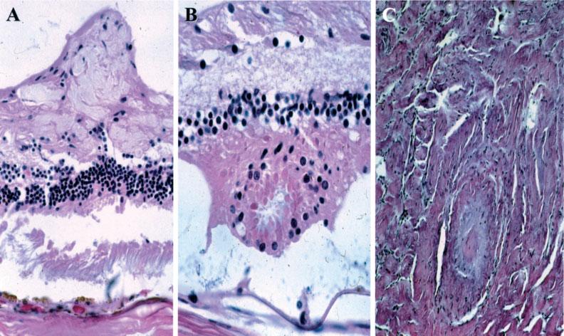
Figure 2.

Other NF2-associated lesions. (A) Retinal tuft arising from the nerve fibre layer. (B) Retinal dysplasia showing a rosette-like configuration. (C) Neurofibroma in the optic nerve (haematoxylin and eosin; original magnification, A, ×200; B, ×400; C, ×200)
