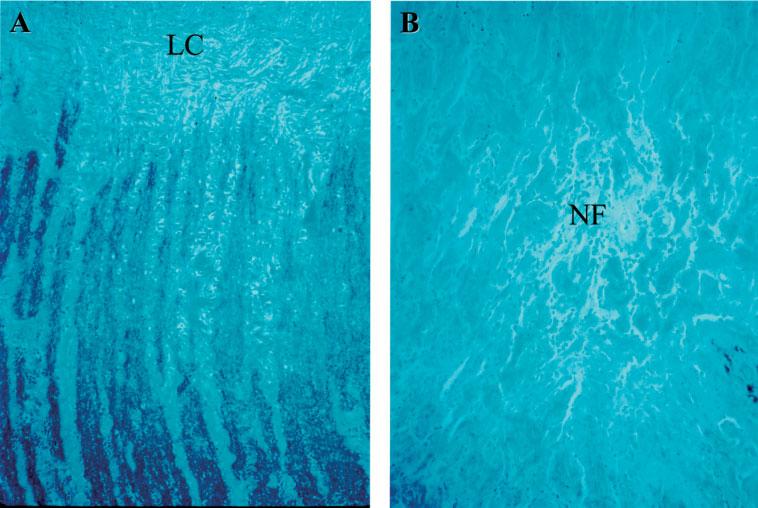
Figure 3.

Expression of merlin in optic nerve. (A) Positive, black colour merlin staining of the optic nerve in an NF2 patient without an optic nerve lesion, showing no difference compared to the normal optic nerve control, where there are Schwann cells and myelinated nerve fibres; note, as expected, absence of staining beyond the level of the lamina cribrosa (LC). (B) Negative merlin staining of the neurofibroma (NF) and positive staining of the optic nerve (left lower corner) of case 4 (immunohistochemical staining against anti-merlin antibody, original magnification, ×100)
