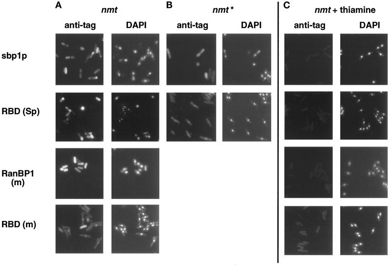
Figure 4.
Localization of exogenous RanBP1 (m), sbp1p, and their RBDs in transformed S. pombe by indirect immunofluorescence. S. pombe cells transformed with full-strength (nmt) or medium-strength (nmt*) plasmids were grown to midlog phase in the presence of thiamine (C) or for an equal amount of time in its absence (A and B). Cells were fixed with paraformaldehyde and permeabilized, and tagged proteins were detected by immunofluorescence using a 1:250 dilution of anti-T7 [(sbp1p, RanBP1 (m), and RBD (m)] or a 1:50 dilution of anti-HA [RBD (Sp)] mAbs, followed by a 1:250 dilution of FITC-labeled anti-mouse secondary antibody. Cells were stained with DAPI before mounting.

