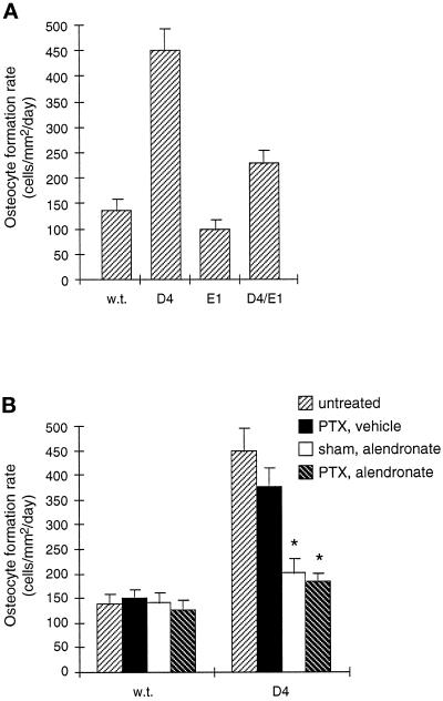
Figure 10.
Osteocyte formation rates in cells/mm2/day in the femoral epiphysis at day 35, calculated as the mathematical product of the epiphyseal osteocyte density (cells/mm3) and the epiphyseal mineral apposition rate (mm3/mm2/day). This index represents the production rate of new osteocytes per unit forming bone surface. Values are shown as mean ± SD. (A) Effects of TGF-β2 overexpression and osteoblastic expression of a truncated type II TGF-β receptor on the epiphyseal osteocyte formation rate. Transgenic E1 mice were crossed to D4 mice to generate double transgenic D4/E1 mice. Although the epiphyseal osteocyte density in D4/E1 mice is at the wild-type level (Figure 3A), the osteocyte formation rate in these mice is partially reduced compared with D4 mice since their epiphyseal mineral apposition rate is still elevated (Figure 7). (B) Effects of alendronate and parathyroidectomy on the epiphyseal osteocyte formation rate. Alendronate treatment with or without parathyroidectomy (∗) dramatically inhibits the increase in osteocyte formation rate in D4 mice because it inhibits both the osteocyte density increase (Figure 4A) and the mineral apposition rate increase (Figure 8) caused by TGF-β2 overexpression.

