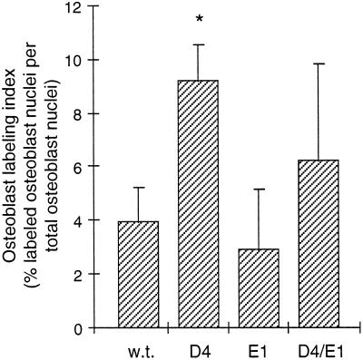
Figure 6.
Kinetics of osteoblast differentiation in the femoral periosteum. Mice were injected with BrdU on day 12 and killed on day 16. The labeling index was calculated as the percentage of labeled periosteal osteoblast nuclei per total periosteal osteoblast nuclei counted in immunostained sections of the femoral diaphysis at the level of the third trochanter. Values represent the mean ± SD of four mice per group. Overexpression of TGF-β2 in D4 mice increased the osteoblast labeling index significantly over wild-type (∗, p < 0.002); the labeling index in E1 mice was not significantly different from wild-type.

