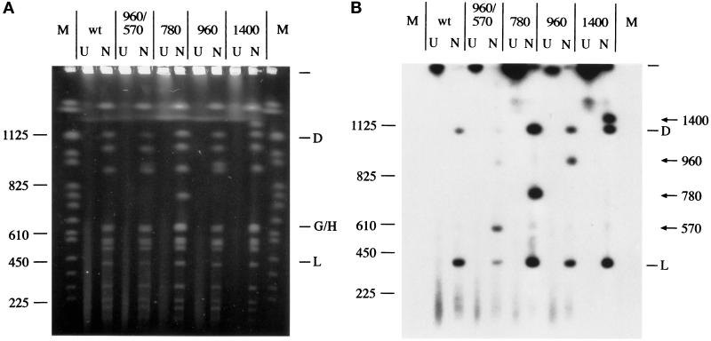
Figure 3.
Pulsed field gel analysis of amplified DNA in strains with sod2 at the gln1 locus. Undigested (U) or NotI-digested (N) total chromosomal DNA samples from wild-type (wt) and four representative sod2-amplified strains were separated by CHEF gel electrophoresis. Lanes are marked with the corresponding size in kilobases of the extra sod2-containing fragments. (A) Ethidium bromide–stained gel. (B) Southern transfer probed with a sod2 probe. NotI restriction fragments D (contains gln1), G/H, and L (contains sod2) are labeled. Arrows denote the amplified sod2 DNA. sod2 hybridization to NotI fragment L in these strains is due to residual sod2 sequences at the normal locus. S. cerevisiae chromosomes in agarose plugs (Life Technologies) were used as DNA size markers (left) and are given in kilobases.

