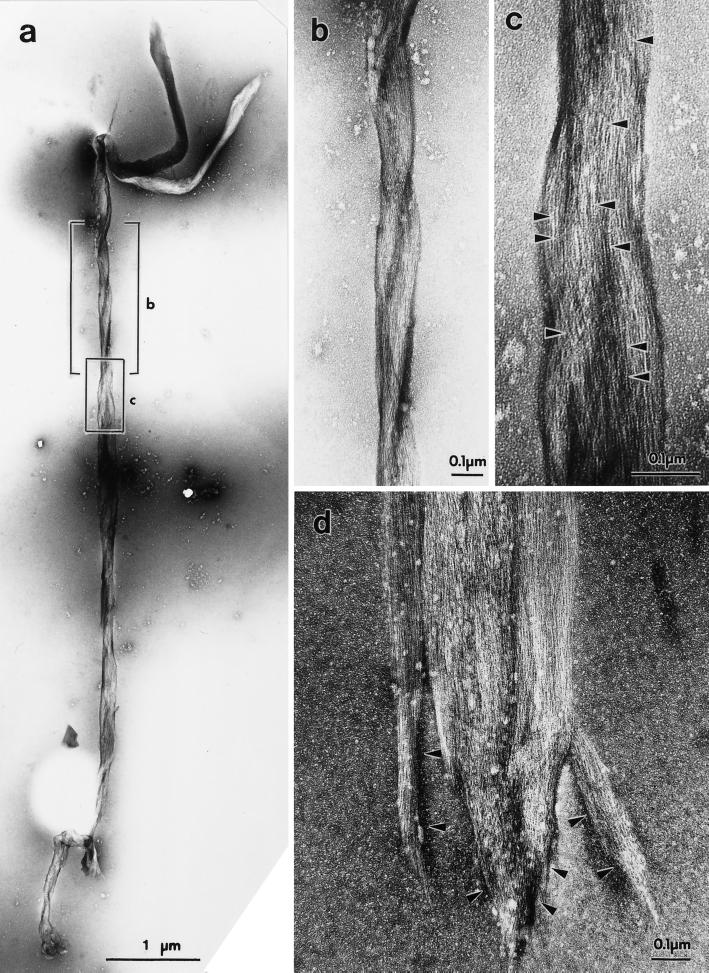
Figure 1.
Electron micrographs of negatively stained stress fibers isolated from bovine endothelial cells (a–c) or human foreskin fibroblasts (d). In a, one whole isolated stress fiber is shown. Two boxed areas in a are enlarged and shown in b and c. Isolated stress fibers consisted of bundles of thin microfilaments. Globular actin monomers in individual filaments are clearly seen (c, arrowheads). Note that the stress fiber in a shows gentle twisting (better shown in b). The ends of isolated stress fibers showed distinct specialization, such as enlargement (a) and finger-like projections (d, arrowheads).

