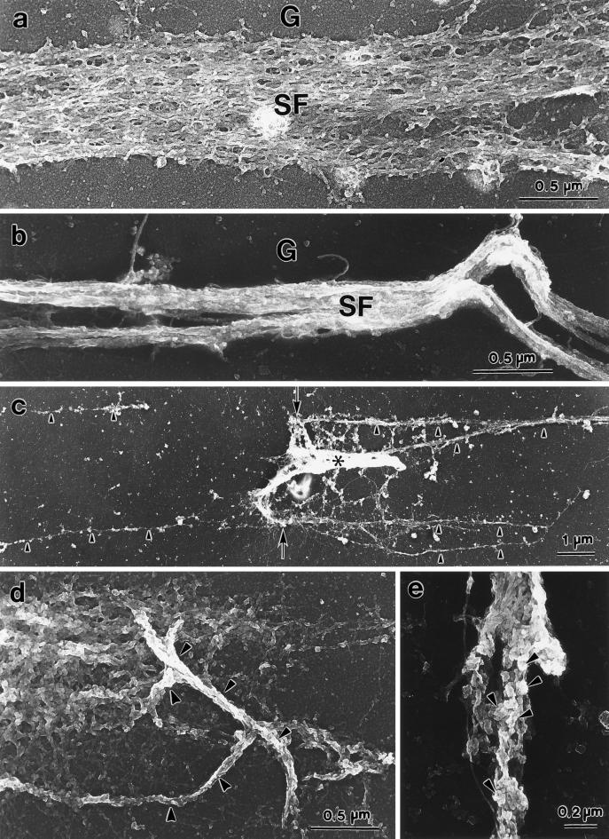
Figure 11.
Replica electron microscopy of isolated stress fibers before and after Mg-ATP treatment. A loose bundle of microfilaments of an isolated stress fiber before Mg-ATP addition is shown (a). Individual microfilaments can be observed. After Mg-ATP addition, microfilaments in a bundle were no longer loose but were tightly packed (b). (c) Low-power view of an attached stress fiber at the end of shortening. The stress fiber (asterisk) is short and dense. It appears to have “contracted” from the left side of the micrograph and flipped over at the end about an imaginary line indicated by the two arrows. Note the extracellular matrix materials on the glass surface (c, arrowheads). Tangled stress fibers (d, arrowheads) and stress fibers exhibiting small, knot-like structures on the surface (e, arrowheads) are illustrated. G, glass surface; SF, stress fiber.

