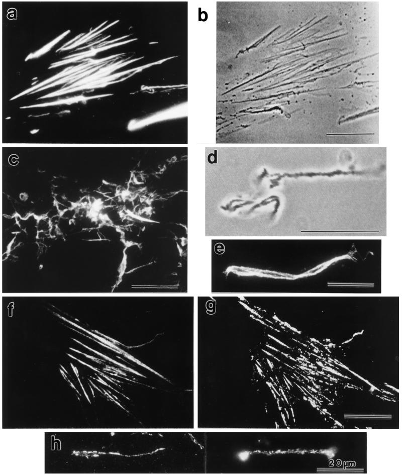
Figure 3.
stress fibers with fluorescein-labeled phalloidin (f) and anti-vinculin (g) is shown. Fractionated stress fibers stained with anti-vinculin are shown (h). Note the dotty anti-vinculin staining pattern along the stress fibers (h). F-actin and vinculin distribution in isolated stress fibers. Attached stress fibers stained with fluorescein-labeled phalloidin (a) and a video-enhanced phase-contrast image of the same sample (b) are shown. Fractionated stress fibers stained with fluorescein-labeled phalloidin (c and e) and a video-enhanced phase-contrast image of a small tangle of isolated stress fibers in the same fraction as in c (d) are shown. Double staining of attached

