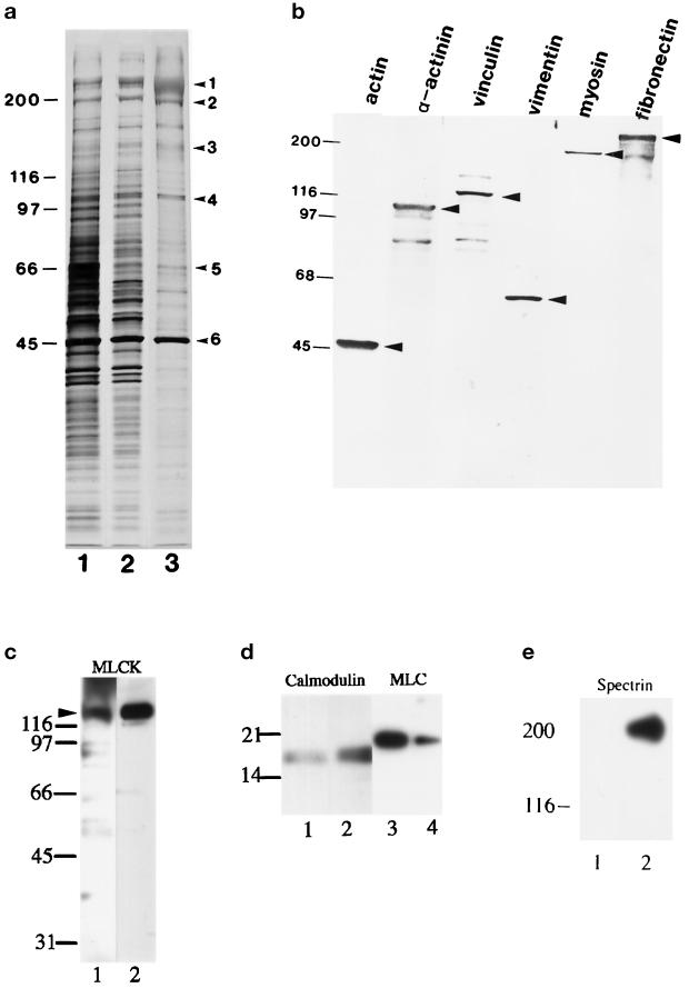
Figure 5.
Gel electrophoresis and immunoblotting of isolated stress fibers. SDS-PAGE gels of a crude extract of fibroblasts (a, lane 1), a TEA-treated cell fraction (a, lane 2), and fractionated stress fibers (a, lane 3) are shown after silver staining. The identity of six major bands (1, fibronectin; 2, myosin; 3, vinculin; 4, α-actinin; 5, vimentin; and 6, actin) in the isolated stress fiber (a, lane 3, arrowheads) was determined by immunoblotting (b). Antibodies used in the immunoblotting analyses in b were anti-α-smooth muscle actin, anti-α-actinin, anti-vinculin, anti-vimentin, anti-myosin, and anti-fibronectin. The presence of myosin light chain kinase (c), calmodulin (d, lanes 1 and 2) and myosin regulatory light chain (d, lanes 3 and 4) was also detected by immunoblotting. Fractionated stress fibers (c, lane 1; d, lanes 1 and 3) and a crude extract of guinea pig aorta as a positive control (c, lane 2; d, lanes 2 and 4) were immunoblotted with anti-myosin light chain kinase (c), anti-calmodulin (d, lanes 1 and 2) and anti-myosin regulatory light chain (d, lanes 3 and 4). Fractionated stress fibers (e, lane 1) and a crude extract of fibroblasts (e, lane 2) were immunoblotted with anti-α-spectrin. Note that no immunoreactivity was detected in the stress fiber sample. Arrowheads in b and c indicate the position of the intact antigen bands.

