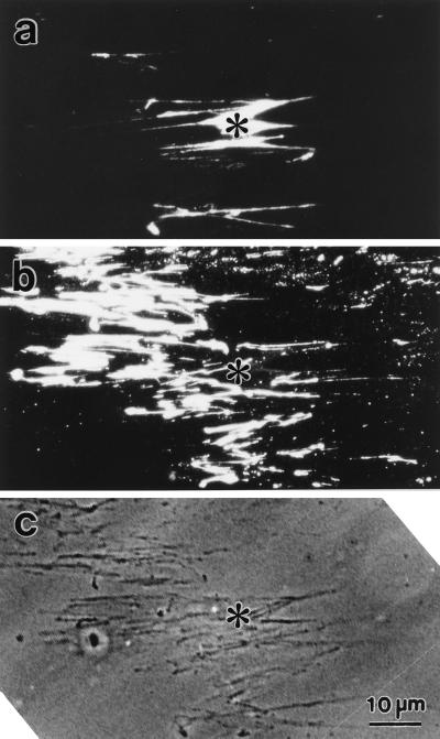
Figure 7.
Identification of the material left behind shortening stress fibers. Attached stress fibers were treated with Mg-ATP, fixed, and stained doubly with rhodamine-labeled phalloidin (a) and anti-fibronectin (b). A video-enhanced phase-contrast image of the same field of view is shown in c. Phalloidin staining shows contracted stress fibers in the center of the micrograph (a), but the fibrous anti-fibronectin pattern occupies a wide portion of the field (b). Note that the anti-fibronectin staining structures are detectable in the phase-contrast micrograph (c). Asterisks indicate the same position in the three micrographs.

