
Fig. 2.
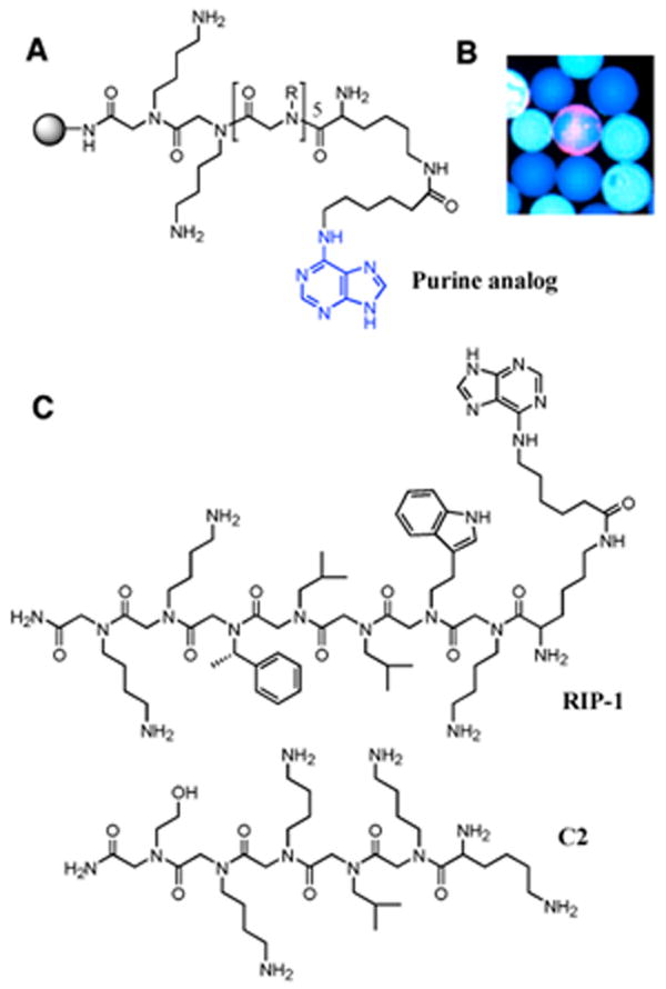
Isolation and characterization of RIP-1. (A) General structure of the purine-capped peptoid library. (B) A fluorescence micrograph of a bead scored as a “hit” (red halo) in the midst of large number of beads scored as negatives (blue). (C) The structure of the hit, RIP-1, as determined by Edman sequencing and C2, a control peptoid not selected for binding to the proteasome.