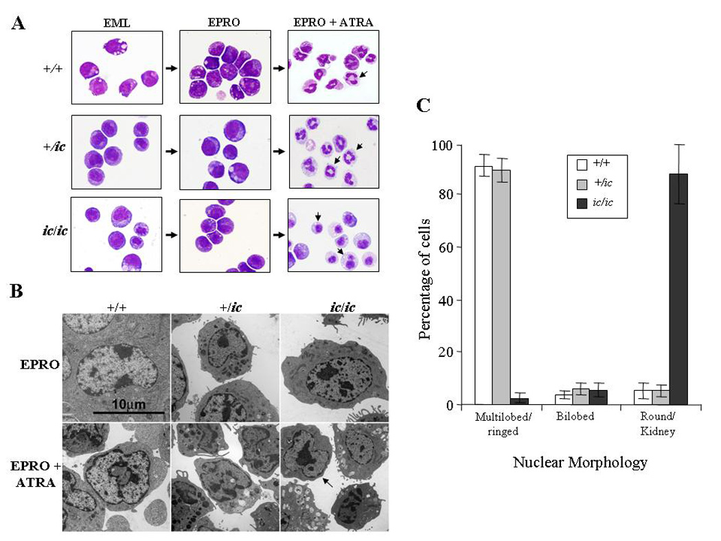
Figure 2. Severe hypolobulation of nuclei in differentiated EPRO-ic/ic cells.

EML-+/+, -+/ic and −ic/ic cells were induced with ATRA (10 µM) plus IL-3 to differentiate into promyelocytic EPRO cells and then further differentiated into mature neutrophils with a second dose of ATRA. (A) Cytospin smears of control +/+ and +/ic vs. ic/ic cells were stained with Wright-Giemsa and then photographed at X 60 magnification using an Olympus BX41 microscope and a DP71 digital camera with accompanying software. Shown are random representations of cells from each stage of development. Arrows in pictures of ATRA-induced EPRO-+/+, -+/ic cells and −ic/ic cells indicate differences in lobulation between each genotype. (B) Electron micrographs of EPRO and ATRA-induced cells from +/+ vs. +/ic and ic/ic cells. (C) Percentages of ATRA-induced EPRO cells with different nuclear morphologies. At least 5 separate inductions of each genotype were performed, and the number of cells exhibiting each type of nuclear morphology was counted at X 40 magnification using an Olympus BX41 microscope. Shown are the average counts of approximately 100 analyzed cells from each induction.
