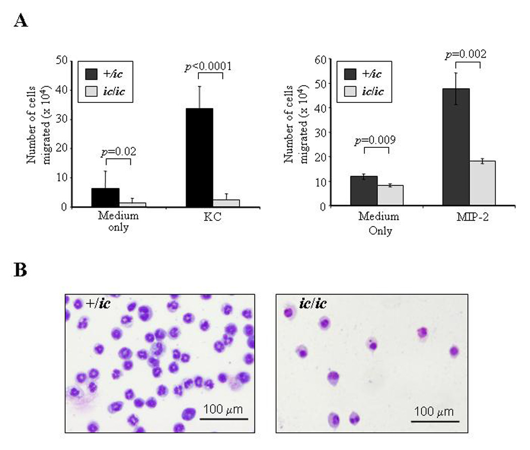
Figure 4. Neutrophil chemotaxis is severely inhibited by the loss of LBR expression.

(A) Chemotaxis in response to KC and MIP-2 is reduced in differentiated EPRO-ic/ic vs. -+/ic cells. ATRA-induced cells were incubated in transwell plates in which each chemoattractant was placed in the lower chamber and cells (1 ×10⁶) were placed in the upper chamber, each separated by a 3.0 µm polycarbonate membrane. The chambers were incubated for 2 hours and the number of cells that migrated into the bottom chamber was counted by trypan blue exclusion. P values for comparisons between genotypes are shown above each bar set. (B) Cells isolated from the bottom chambers after KC-induced chemotaxis were Wright-Giemsa-stained and photographed as described in Figure 2. Shown is a representation of the differentiated cells that migrated after 2 hours of incubation.
