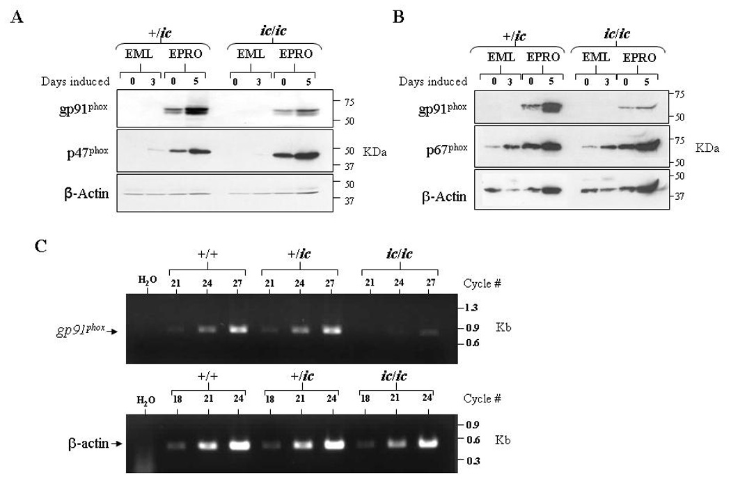
Figure 6. Loss of LBR expression inhibits gp91phox expression in differentiated EPRO-ic/ic cells.

Western blots were generated from uninduced and induced +/ic and ic/ic cells, and then sequentially probed with antibodies for (A) gp91phox and p47phox or (B) gp91phox and p67phox. Both blots were also probed for β-actin expression, and equivalent, total numbers of cells were used for each lysate. (C) Semi-quantitative RT-PCR analysis reveals reduced gp91phox transcript expression in ATRA-induced EPRO-ic/ic as compared to +/+ and +/ic cells. Shown are ethidium bromide-stained gels containing RT-PCR amplified products from each genotype using increasing numbers of cycles, using primers specific to mouse gp91phox (top) and mouse β-actin (bottom) as a loading control.
