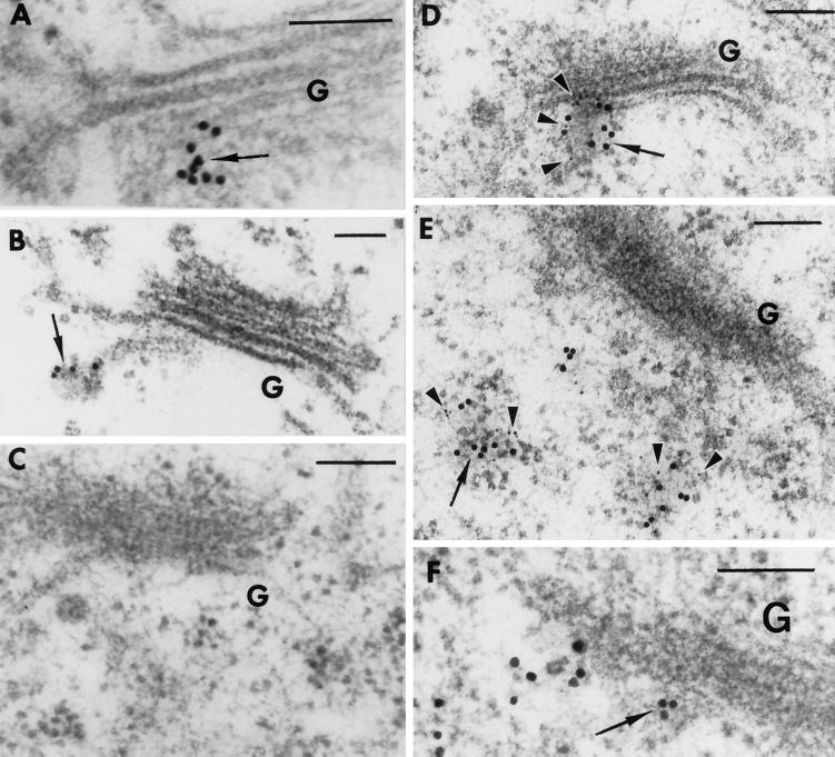
Figure 8.
In situ localization of T7-AtVTI1a and AtELP on ultrathin sections of Arabidopsis roots from T7-AtVTI1a transgenic plants. T7-AtVTI1a and AtELP are localized on the TGN and on dense vesicles. (A and B) Ultrathin sections were incubated with T7 mAb followed by rabbit anti-mouse IgG and biotinylated goat anti-rabbit secondary antibody and were visualized with streptavidin conjugated to 10-nm colloidal gold. (C) Control. The ultrathin sections were treated with the same procedure as described in A and B, except T7 mAb was substituted with 2% BSA in PBS. T7-AtVTI1a and AtELP are colocalized on the TGN (D) and on dense vesicles (E). (D and E) Ultrathin sections were incubated with T7 mAb followed by rabbit anti-mouse IgG and biotinylated goat anti-rabbit secondary antibody and were visualized with streptavidin conjugated to 10-nm colloidal gold. After the second fixation step (see MATERIALS AND METHODS), the same sections were incubated with antiserum to AtELP, followed by biotinylated goat anti-rabbit secondary antibody, and then visualized with streptavidin conjugated to 5-nm colloidal gold. (F) Control section. The same procedures were used as in D and E, except preimmune serum was used in the place of AtELP antiserum. G, Golgi; arrow, AtVTI1a; arrowhead, AtELP; bar, 0.1 μm.

