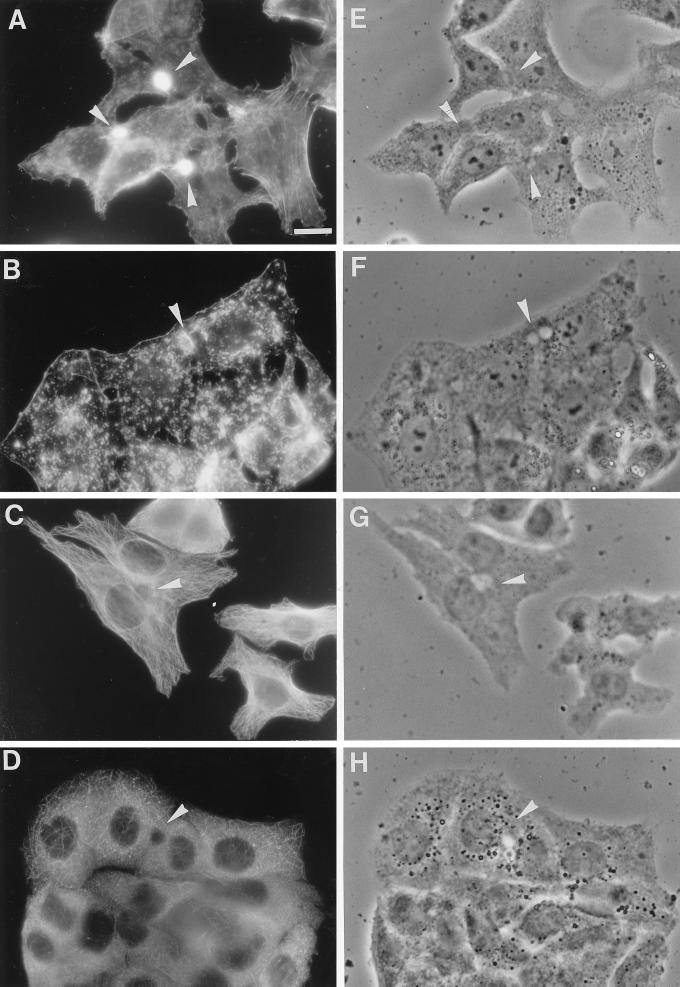
Figure 1.
Effect of cytoskeletal drugs on the distribution of actin filaments and microtubules in HepG2 cells. The effect of cytD on actin filaments was determined by incubating the cells with 10 μg/ml cytD in HBSS for 30 min at 37°C. After the incubation, the cells were washed and fixed in ethanol for 10 s at −20°C. Actin filaments were stained with 100 ng/ml TRITC-labeled phalloidin in PBS for 30 min at room temperature. Note the extensive staining of actin filaments around the bile canaliculus (arrowhead) and the additional staining at the basolateral membrane in control cells (A), whereas in cytD-treated cells (B) mainly small aggregates and a strongly reduced labeling around the bile canaliculus are observed. By staining cells with anti-β-tubulin, the effect of nocodazole on microtubules was evaluated. Cells were preincubated with 33 μM nocodazole in HBSS for 2 h at 37°C. The cells were then fixed with 3% paraformaldehyde for 30 min at room temperature and permeabilized with 0.1% Triton X-100. Indirect immunofluorescence staining with an antibody against β-tubulin revealed that in control cells, the microtubules formed a delicate network (C) that was severely disrupted after nocodazole treatment (D). (E–H) Phase-contrast images of A–D, respectively. Bar, 10 μm.

