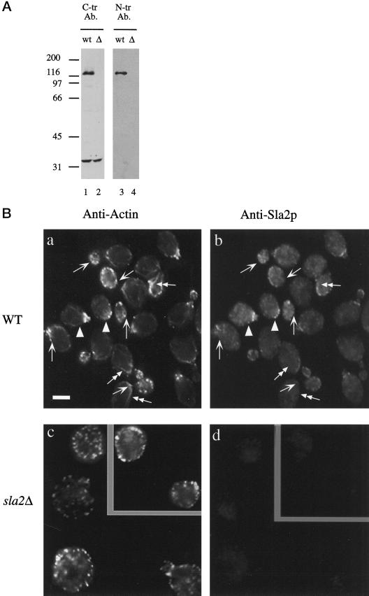
Figure 2.
Subcellular localization of Sla2p. (A) Affinity-purified anti-Sla2p antibodies recognize a 116-kDa protein species in wild-type yeast whole-cell extracts. Lanes 1 and 2, immunoblot using the affinity-purified antibody against the C terminus of Sla2p. Lanes 3 and 4, immunoblot using affinity-purified antibody against the N terminus of Sla2p. Lanes 1 and 3 contain whole-cell extract from wild-type cells. Lanes 2 and 4 contain whole-cell extract from sla2Δ cells. (B) Indirect immunolocalization shows that Sla2p localizes to cortical structures the yeast cell. This localization largely, but not completely, overlaps with that of cortical actin. Arrows indicate patches that contain actin but no detectable Sla2p. Arrowheads show patches that contain Sla2p but no detectable actin. Double-headed arrows indicate actin cables, which contain no Sla2p. Diploid wild-type (DDY288; a and b) and sla2Δ cells (DDY540; c and d) were processed for immunofluorescence using affinity-purified antibody against the C terminus of Sla2p and anti-yeast actin antibodies, as described in MATERIALS AND METHODS. (a and c) Fluorescein staining of actin structures. (b and d) CY3 staining of Sla2p structures.

