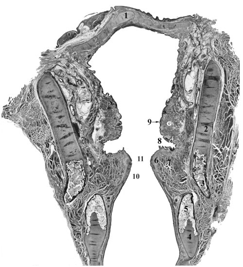
Figure 3.
Coronal section of the larynx from 56 year old female illustrating major structural landmarks. Safran-Hematoxylin Stain, 2.5x original magnification. 1 – epiglottis, 2 – unossified thyroid cartilage, 3 – ossified portion of thyroid cartilage, 4 – unossified cricoid cartilage, 5 – ossified portion of cricoid cartilage, 6 – lamina propria of vocal fold, 7 – thyroarytenoid muscle (the major constrictor of vocal fold), 8 – laryngeal ventricle (lined of glandular epithelium), 9 – vestibular fold with G – mucous-serous glands, 10 – conus elasticus, 11 – glottal space.

