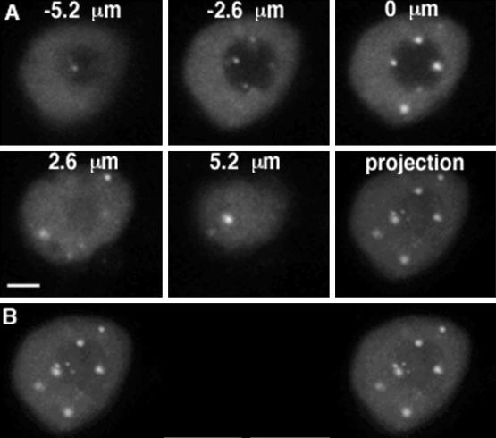
Figure 2.
Confocal fluorescence microscopy of U2B"-GFP in a living BY-2 cell. (A) Single confocal optical sections of a BY-2 cell nucleus are shown, with 2.6 μm between consecutive sections, followed by a projection of the 3-D data stack. Coiled bodies can be found in the nucleoplasm and nucleolus, but most frequently just inside the nucleolar periphery. (B) Stereo projection of the confocal series shown in panel A. Bar, 5 μm.

