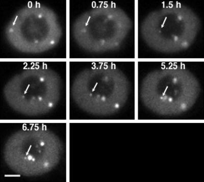
Figure 3.
Time-lapse confocal microscopy of U2B"-GFP in living BY-2 cells. Projections of three or four confocal optical sections are shown at each time point. Coiled body marked with an arrow demonstrates that coiled bodies can move around in the nucleoplasm and nucleolus, and often move along the periphery of the nucleolus. Other, unmarked coiled bodies show smaller movements. Bar, 5 μm.

