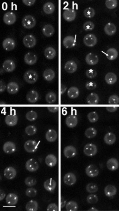
Figure 6.
Time-lapse confocal microscopy of U2B"-GFP in stable Arabidopsis transformants. Projections of series of confocal optical sections through the root epidermis are shown at each time point. Many coiled bodies are clearly mobile. Several coiled bodies coalesce (arrows). Two cells underwent division during this sequence, and the newly divided cells decreased their coiled body numbers quickly (asterisk). Bar, 10 μm.

