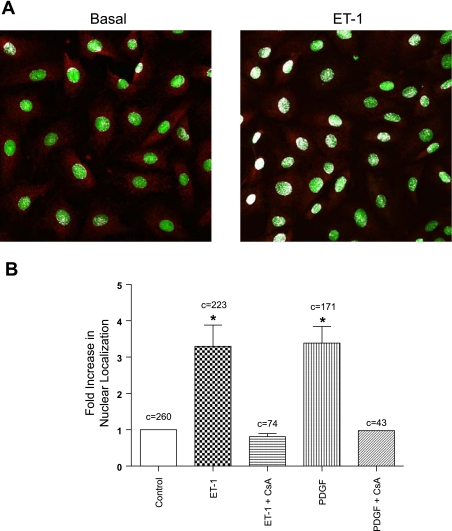
Fig. 2.
Stimulation of NFATc3 nuclear translocation in UBSM. A: example micrographs showing the subcellular distribution pattern of NFATc3 under basal conditions and after stimulation with endothelin-1 (ET-1). B: treatment with either ET-1 or platelet-derived growth factor (PDGF) led to a significant increase in NFATc3 nuclear localization compared with basal conditions (*P < 0.05; c = number of cells analyzed). Treatment with FK506 and cyclosporine A (CsA, 1 μM; 30 min) before the addition of the PDGF or ET-1 blocked NFATc3 nuclear translocation.

