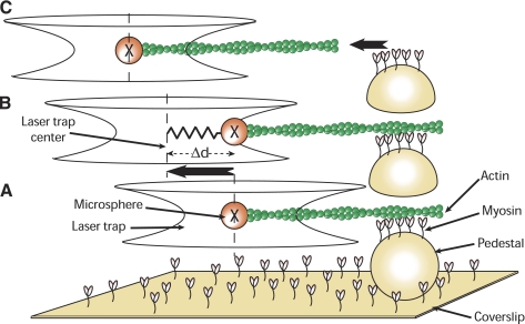
Fig. 1.
A: a single beam laser trap is used to capture a 3-μm-diameter microsphere coated with N-ethylmaleimide-modified myosin. A tetramethylrhodamine isothiocyanate (TRITC)-labeled actin filament is attached to the microsphere and brought in contact with unphosphorylated myosin randomly adhered to a 4.5-μm pedestal on a coverslip. B: after time is allowed for the binding of unphosphorylated myosin to actin, the laser trap is moved away from the pedestal at constant velocity. Initially, the microsphere remains at the same position, now offset from the center of the trap. C: when the pulling force exerted by the trap exceeds the binding force of the unphosphorylated myosin molecules, the microsphere springs back into the trap center, its unloaded position. Unbinding force (Funb) is equal to the product of the maximal distance between the bead and trap center [maximal displacement (Δd)] by the trap stiffness [calibrated using the Stokes force method (7)].

