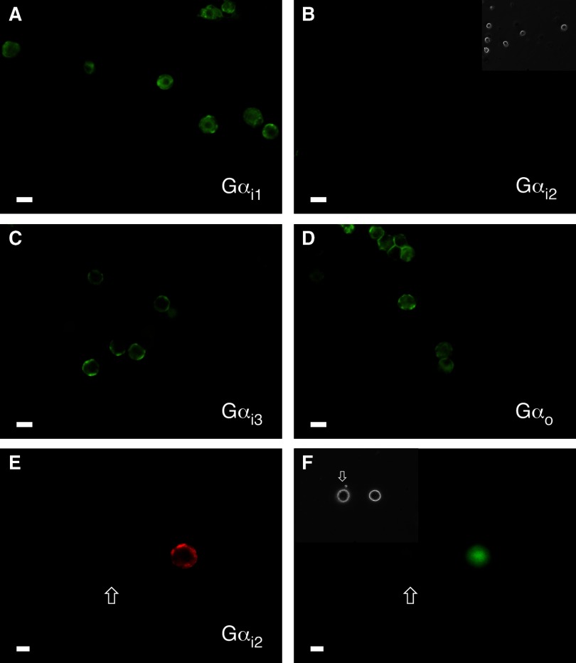
FIG. 1.
Immunofluorescence imaging reveals stellate ganglion (SG) neurons express pertussis toxin (PTX)-sensitive Gαi1, Gαi3, and Gαo but not Gαi2 subunits. Shown are fluorescence images of SG neurons fixed, permeabilized, and stained with primary antibodies against Gαi1 (A), Gαi2 (B), Gαi3 (C), and Gαo (D) followed by Alexa Fluor 488-conjugated IgG secondary antibody. The neurons were imaged at 20 × with a filter set containing an excitation filter at 480 ± 15 nm, a dichroic beam splitter of 505 nm (LP) and an emission filter at 535 ± 20 nm. The images were pseudocolored; scale bars represents 20 μm. B, inset: the phase image for the corresponding fluorescence field shown. As positive controls for Gαi2 staining, fluorescence images of SG neurons transfected with Gαi2 and enhanced green fluorescent protein (pEGFP) cDNA constructs are shown in E and F. The secondary antibody employed in E was Alexa Fluor 568 and imaged with a filter set containing an excitation filter at 540 ± 15 nm, a dichroic beam splitter of 585 nm (LP) and an emission filter at 620 ± 30 nm. F, inset: the phase image of the corresponding fluorescence field. Scale bars represent 20 μm. Arrow in all 3 images points to non-Gαi2- and GFP-expressing neuron.

