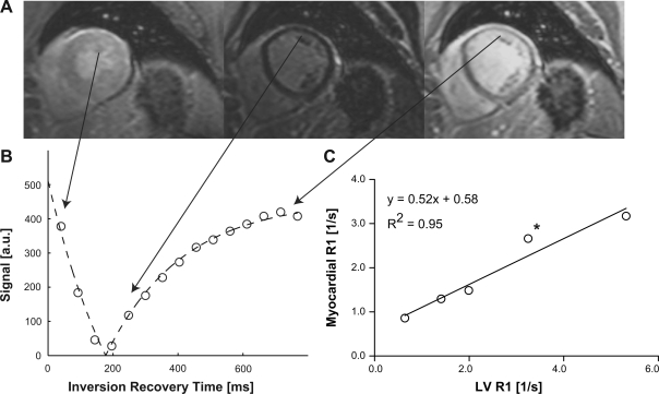
Fig. 1.
A: a series of inversion recovery images was acquired in a female patient diagnosed with idiopathic dilated cardiomyopathy (IDC) [ejection fraction = 45%; end-diastolic volume (EDV) indexed by height = 96 ml/m] with a cardiac and respiratory-gated Look-Locker sequence with up to 30 phases, using a segmented gradient-echo readout with a train of low-angle (12°) excitation pulses. Only 3 out of 15 images are shown here and in order of increasing inversion times from left to right. B: the graph shows the measured signal intensity in 1 of 8 transmural wall segments (open circles) against the time delay after the nonslice-selective inversion pulse. The continuous lines represent least-squares fits to the data points, using a model equation for the inversion-recovery measured from magnitude images. C: the relaxation rate (R1 = 1/T1) in tissue was plotted against the corresponding R1 in the left ventricular (LV) blood pool. The partition coefficient was determined by linear regression and was 0.52 ± 0.07 (mean ± SE) in this patient. The asterisk denotes the measurement corresponding to the images in A and the graph in B. a.u., Arbitrary units.

