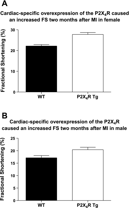
Fig. 3.
The increase in LV basal FS in P2X4R Tg mice was sustained at 2 mo after infarction in both sexes. Two-dimensionally directed M-mode echocardiography was carried out as described in methods. The basal values for LV FS of 22 WT and 17 Tg female (A) and of 23 WT and 15 Tg male (B) were summarized as means ± SE. P2X4R Tg mice showed significantly greater FS than did the WT mice (P < 0.05, unpaired t-test).

