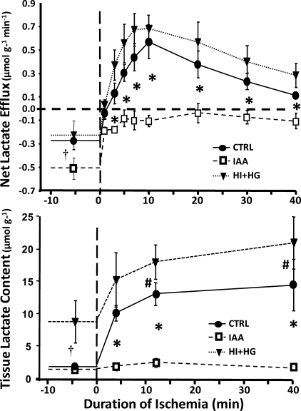
Fig. 3.
Net myocardial lactate uptake (top) and myocardial lactate concentration (bottom) plotted as a function of time in the CTRL, IAA, and HI + HG groups under normal flow conditions and during 40 min with a 60% decrease coronary blood flow. *P < 0.01 for IAA vs. both CTRL and HI + HG at given time point; †P < 0.05 for HI + HG vs. both CTRL and IAA at preischemic baseline; #P < 0.05 for HI + HG vs. CTRL at given time point.

