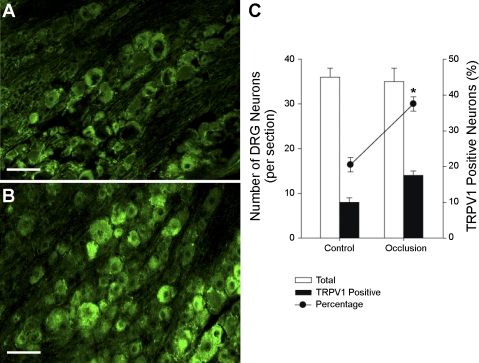
Fig. 1.
Transient receptor potential vanilloid type 1 (TRPV1) receptor expression in the dorsal root ganglion (DRG) (L4–L6) neurons. Photographs show TRPV1 immunoreactivity in the DRG from a control rat (A) and a rat with 24 h arterial occlusion (B). The receptors were seen in small and medium diameter of cells and rarely in large diameter of cells. Scale bar = 35 μm. The percentage of TRPV1-positive neurons was significantly higher in rats with arterial occlusion (n = 5) than that in sham-operated control rats (n = 5) (C). *P < 0.05 compared with control group.

