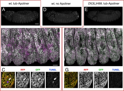
Fig. 4.
Comparing Apoliner and TUNEL in normal and apoptosis-deficient Drosophila embryos. (A–C) TUNEL and Apoliner in a control (+/+ or H99/+) embryo. (A) TUNEL signal as seen in a lateral view (projection of 16 consecutive confocal slices). (B) Apoliner in the embryo shown in A, with RFP displayed in magenta and GFP in green, resulting in a white signal upon colocalization. Again, 16 confocal slices are shown, but this time they are projected using the 3D function of Volocity (Improvision). (C) High-magnification view of one confocal slice from B, showing RFP, GFP, and TUNEL, and the overlay on the left-hand side. The arrow points to a shrunken Apoliner-positive cell, which is also positive for TUNEL, an indication of advanced apoptosis. At this late stage, the GFP signal is no longer detectable, presumably because of the high proteolytic activity in this cell (see Fig. S1). (D) TUNEL staining in a wild-type (or H99/+) embryo that does not express Apoliner. Note that the number of TUNEL-positive nuclei is approximately the same as in A, indicating that Apoliner does not interfere significantly with developmental apoptosis. (E–G) TUNEL and Apoliner in a H99-deficient embryo. (E) H99-deficient embryo stained for TUNEL; very little staining can be detected. (F) Apoliner in the embryos shown in E, with RFP in magenta and GFP in green (again, 16 confocal slices projected using the 3D function in Volocity–Improvision). (G) High-magnification view of one confocal slice from F (H99-deficient embryo). In the trunk epidermis, no TUNEL signal or Apoliner cleavage can be detected, whereas an occasional signal is observed in the head region (data not shown; also see ref. 8). [Scale bar for A, D, and E (shown in A), 50 μm; scale bar for B and F (shown in B), 10 μm.]

