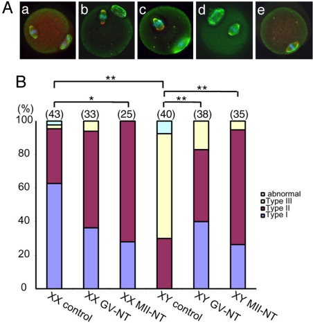
Fig. 1.
Spindle microtubules in the MII oocytes after in vitro maturation (IVM), with or without nuclear transfer. (A) Immunolabeling of α-tubulin (green) and γ-tubulin (red) counterstained with DAPI (blue). (a) Type I spindle found in the XX control group. α-tubulin labeling shows a barrel-shaped microtubule spindle in a parallel position to the oolemma. γ-tubulin labeling is seen in punctuate foci at both poles. The condensed chromosomes are aligned along the midzone. The first polar body is seen on the bottom right. (b) Type II spindle found in the XY control group. The microtubule spindle shows globally normal shape, but oriented in a perpendicular position to the oolemma. Multiple γ-tubulin foci are spread over both poles, which are considerably wider that the poles of Type I spindle. The condensed chromosomes are loosely aligned at the midzone. The first polar body is seen on the top. (c) Type III spindle found in the XY control group. The microtubule spindle is bulky, i.e., one pole is wider than the midzone, and oriented in a perpendicular position to the oolemma. Multiple γ-tubulin foci are spread over the spindle. The condensed chromosomes are scattered around the midzone. The first polar body is seen on the bottom right. (d) Type I spindle observed in the XY GV-NT group. The microtubule spindle has normal shape with 1 or 2 γ-tubulin foci at each pole. The condensed chromosomes are aligned at the midzone. The first polar body is seen on the top left. (e) Type I spindle observed in the XY MII-NT group. A barrel-shaped microtubule spindle is oriented in a parallel position to the oolemma. Two γ-tubulin foci are seen at each pole. The condensed chromosomes are aligned at the midzone. The first polar body has been removed during nuclear transfer. (B) Percentages of MII oocytes containing each type of meiotic spindles. The total number of oocytes examined is indicated on the top of each column. *, P < 0.01; **, P < 0.001 by χ2 test. Type I, II, and III spindles are defined above. The “abnormal” spindle is defused or contains tripolar microtubule spindles.

