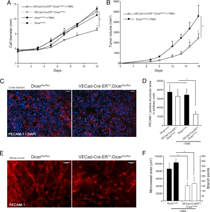
Fig. 3.
Endothelial Dicer inactivation reduces tumor angiogenesis and progression. (A and B) LLC cells were implanted intramuscularly into the calves of 8-week-old Dicerflox/flox or VECad-Cre-ERT2;Dicerflox/flox mice (A) or implanted s.c. in the dorsal flank of Dicerflox/flox and TMX-treated VECad-Cre-ERT2;DicerΔ/Δ mice (B), and tumor growth was assessed. n = 4 per group. P < 0.05 using a two-way ANOVA, followed by post hoc Bonferroni test. (C) Representative PECAM-1 staining (red) of cross-sections from tumors showing reduced tumor angiogenesis in VECad-Cre-ERT2;Dicerflox/flox mice. Tissues were counterstained with DAPI (blue). (D) Quantification of PECAM-1-positive structures in the tumors of the indicated groups of mice. For each tumor, two to four images from three sections from two different tumor pieces were quantified. (E) Representative whole-mount PECAM-1 staining from tumors isolated from dorsal flank showing reduced tumor-induced angiogenesis in VECad-Cre-ERT2;Dicerflox/flox mice. (F) Quantification of microvessel area and branch points, left and right bars for each group, respectively; data are from three to five images per sample. *, P < 0.05, compared with Dicerflox/flox by Student's t test. All data are mean ± SEM. (Scale bars: C and E, 100 μm.)

