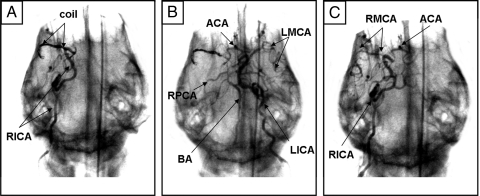
Fig. 2.
C-arm fluoroscopy enabled real-time verification of MCA occlusion and reperfusion in the canine brain. (A) The matrix coil was deployed into the RMCA, occluding the M1 segment, as confirmed by RICA contrast injection. (B) LICA angiography demonstrated that the LMCA was still intact, along with the ACA, BA, and RPCA. The RMCA still did not fill. (C) After 1-h occlusion, the matrix coil was retrieved and RICA arteriogram confirmed that the RMCA had reperfused.

