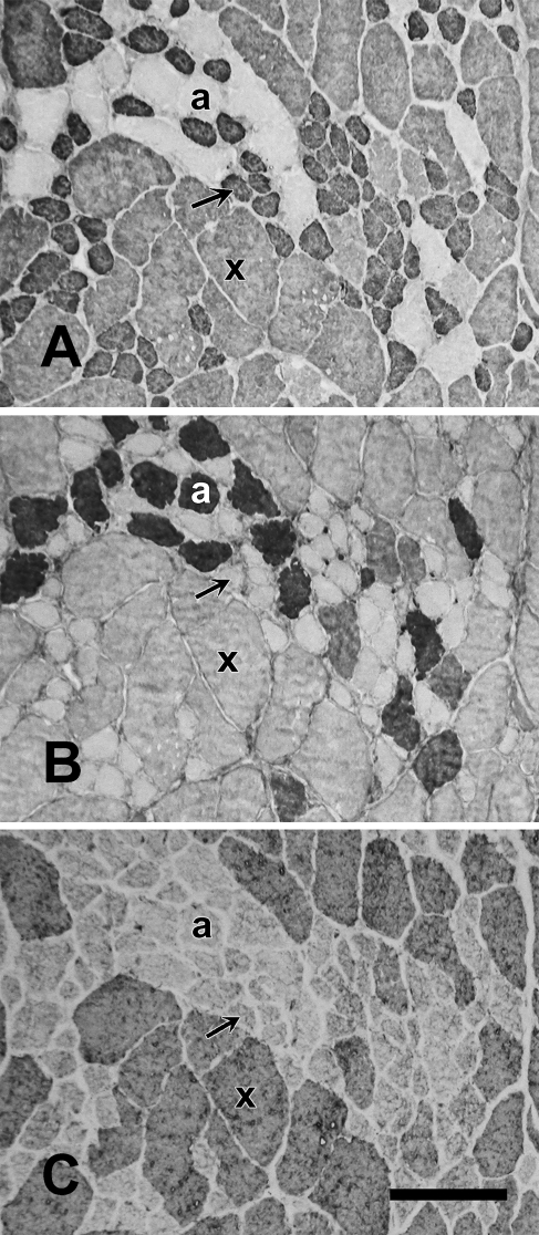
Figure 11.
High-power photomicrograph of immunoperoxidase-stained serial sections of the baboon Ta-V muscle reacting to anti-slow MAb (A), anti-2A MAb (B), and anti-2X MAb (C). Slow fibers (arrow), 2a fibers (a), and 2X fibers (x) are labeled. The anti-2A and anti-2X MAbs are specific, but the anti-slow MAb can be seen to faintly stain 2X fibers that are visibly distinct in fiber size. This cross-reaction was accounted for in quantitative analysis. Bar = 100 mm.

