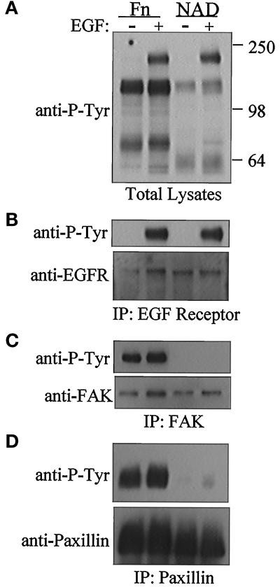
Figure 2.
Cell adhesion and EGF stimulation trigger distinct initial tyrosine phosphorylation events in ECV304 cells. Confluent cells were serum starved for 6 to 8 h, trypsinized, resuspended in 2% BSA, and rocked in suspension for 1 h. Tyrosine phosphorylation levels were measured in cells either replated on fibronectin-coated dishes (Fn) or maintained in nonadherent suspension culture (NAD) for 2 h. Where indicated (+) EGF (1 ng/ml) was added to medium 5 min before cell harvest. Samples were Western blotted with a monoclonal anti-phosphotyrosine antibody and then reprobed with an antibody to the protein of interest. (A) Total cell lysates; blotted with anti-P-tyr. (B) lysates immunoprecipitated with a monoclonal anti-EGF receptor antibody; blotted with anti-P-tyr, reprobed with anti-EGFR. (C) Immunoprecipitation with an anti-FAK antibody; blotted with anti-P-tyr, reprobed with anti-FAK. (D) Immunoprecipitation with an anti-paxillin antibody, blotted with anti-P-tyr, reprobed with anti-paxillin.

