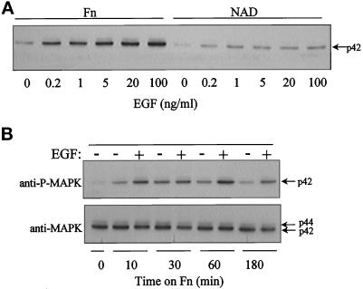
Figure 4.
Anchorage dependence of EGF activation of MAPK. Confluent ECV304 cells were serum starved for 6 to 8 h, trypsinized, and suspended in medium containing 2% BSA. Following a 1-h period in suspension, cells were allowed to adhere to dishes coated with fibronectin or were held in suspension. Where indicated, EGF was added to cells 5 min before harvest. Cells were lysed and analyzed by Western blot analysis for MAPK activation. (A) EGF dose response. Cells were treated with EGF (0–100 ng/ml) after attachment to fibronectin (Fn), or after suspension culture (NAD), for 3 h. Lysates were probed with an anti-active MAPK antibody. (B) Time course. Cells were replated on a fibronectin substratum for various periods of time and then treated with 1 ng/ml EGF (+) or not (−). Top panel, anti-active MAPK Western blot. Bottom panel, anti-MAPK Western blot.

