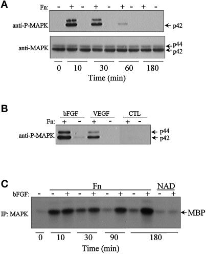
Figure 5.
Mitogen-mediated activation of MAPK is anchorage dependent in primary human endothelial cells (HUVECs). Confluent HUVECs were serum starved for 8 h, trypsinized, and suspended in medium containing 2% BSA. Following a 1-h period in suspension, cells were allowed to adhere to dishes coated with fibronectin (Fn) or held in suspension (NAD). In some cases, the cells were treated for 5 min with bFGF or VEGF. Cells were washed and lysed, and lysates were analyzed for MAPK activation. (A) Cells were maintained in suspension (−) or attached to fibronectin (+) for various intervals, in the absence of growth factors, and the cell lysates were probed with an anti-active MAPK antibody. (B) Cells were replated on fibronectin (+) or held in suspension (−) for 3 h, and then stimulated with either VEGF (20 ng/ml) or bFGF (10 ng/ml) or not stimulated (CTL); cell lysates were probed with an anti-active MAPK antibody. (C) Cells were replated on fibronectin (Fn) or held in suspension (NAD) for 3 h, and then stimulated (+) with bFGF (10 ng/ml) or not stimulated (−); the cell lysates were analyzed using an in vitro MAPK assay, as described in MATERIALS AND METHODS. The autoradiogram represents the 32P phosphorylation of MBP.

