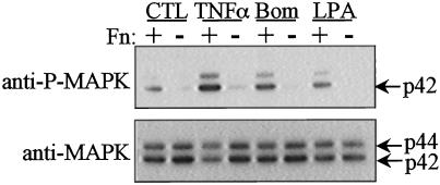
Figure 6.
G protein-coupled receptor agonists and TNF-α show anchorage-dependent activation of MAPK in HUVECs. Confluent HUVECs were serum starved, trypsinized, and suspended in medium containing 2% BSA. Following a 1-h period in suspension, cells were allowed to adhere to dishes coated with fibronectin (Fn) or held in suspension (NAD) for 3 h prior to agonist stimulation. Cells were stimulated with either bombesin (Bom, 5 min, 10 nM), LPA (5 min, 2 μM), or TNF-α (10 min, 10 ng/ml), and lysates were analyzed by Western blotting. The top panel shows the activated forms of MAPK using an anti-active MAPK antibody; the bottom panel shows loading using an anti-MAPK antibody.

